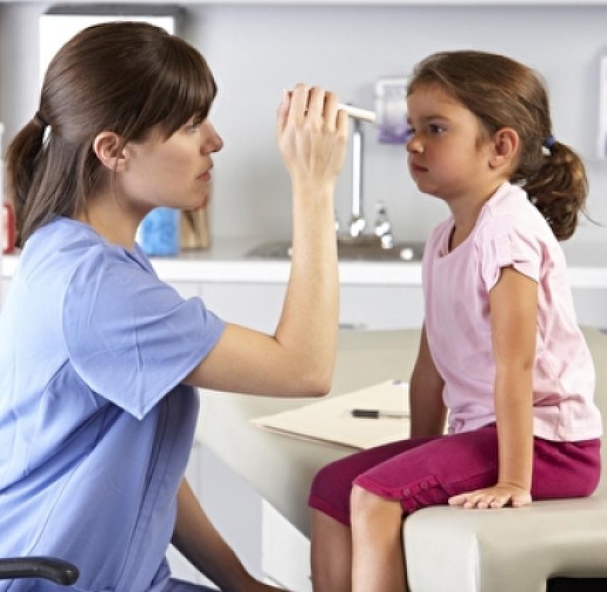
Although the majority of TBIs are mild they can still have serious health implications. Of greatest concern are injuries that can quickly grow worse. All TBIs require immediate assessment by a professional who has experience evaluating head injuries. A neurological exam will assess motor and sensory skills and the functioning of one or more cranial nerves. It will also test hearing and speech, coordination and balance, mental status, and changes in mood or behavior, among other abilities. Screening tools for coaches and athletic trainers can identify the most concerning concussions for medical evaluation.
Initial assessments may rely on standardized instruments such as the Acute Concussion Evaluation (ACE) form from the Centers for Disease Control and Prevention or the Sport Concussion Assessment Tool 2, which provide a systematic way to assess a person who has suffered a mild TBI. Reviewers collect information about the characteristics of the injury, the presence of amnesia (loss of memory) and/or seizures, as well as the presence of physical, cognitive, emotional, and sleep-related symptoms. The ACE is also used to track symptom recovery over time. It also takes into account risk factors (including concussion, headache, and psychiatric history) that can impact how long it takes to recover from a TBI.
When necessary, medical providers will use brain scans to evaluate the extent of the primary brain injuries and determine if surgery will be needed to help repair any damage to the brain. The need for imaging is based on a physical examination by a doctor and a person’s symptoms.
Computed tomography (CT) is the most common imaging technology used to assess people with suspected moderate to severe TBI. CT scans create a series of cross-sectional x-ray images of the skull and brain and can show fractures, hemorrhage, hematomas, hydrocephalus, contusions, and brain tissue swelling. CT scans are often used to assess the damage of a TBI in emergency room settings.
Magnetic resonance imaging (MRI) may be used after the initial assessment and treatment as it is a more sensitive test and picks up subtle changes in the brain that the CT scan might have missed.
Unlike moderate or severe TBI, milder TBI may not involve obvious signs of damage (hematomas, skull fracture, or contusion) that can be identified with current neuroimaging. Instead, much of what is believed to occur to the brain following mild TBI happens at the cellular level. Significant advances have been made in the last decade to image milder TBI damage. For example, diffusion tensor imaging (DTI) can image white matter tracts, more sensitive tests like fluid-attenuated inversion recovery (FLAIR) can detect small areas of damage, and susceptibility-weighted imaging very sensitively identifies bleeding. Despite these improvements, currently available imaging technologies, blood tests, and other measures remain inadequate for detecting these changes in a way that is helpful for diagnosing the mild concussive injuries.
Neuropsychological tests to gauge brain functioning are often used in conjunction with imaging in people who have suffered mild TBI. Such tests involve performing specific cognitive tasks that help assess memory, concentration, information processing, executive functioning, reaction time, and problem-solving. The Glasgow Coma Scale is the most widely used tool for assessing the level of consciousness after TBI. The standardized 15-point test measures a person’s ability to open his or her eyes and respond to spoken questions or physical prompts for movement. A total score of 3-8 indicates a severe head injury; 9-12 indicates moderate injury; and 13-15 is classified as mild injury. (For more information about the scale, see glasgowcomascale.org).
Many athletic organizations recommend establishing a baseline picture of an athlete’s brain function at the beginning of each season, ideally before any head injuries have occurred. Baseline testing should begin as soon as a child begins a competitive sport. Brain function tests yield information about an individual’s memory, attention, and ability to concentrate and solve problems. Brain function tests can be repeated at regular intervals (every 1 to 2 years) and also after a suspected concussion. The results may help health care providers identify any effects from an injury and allow them make more informed decisions about whether a person is ready to return to their normal activities.
Source: Traumatic Brain Injury Hope Through Research, NINDS, Publication date September 2015. http://www.ninds.nih.gov
NIH Publication No. 15-2478
Prepared by:
Office of Communications and Public Liaison
National Institute of Neurological Disorders and Stroke
National Institutes of Health
Bethesda, MD 20892
NINDS health-related material is provided for information purposes only and does not necessarily represent endorsement by or an official position of the National Institute of Neurological Disorders and Stroke or any other Federal agency. Advice on the treatment or care of an individual patient should be obtained through consultation with a physician who has examined that patient or is familiar with that patient's medical history.
All NINDS-prepared information is in the public domain and may be freely copied. Credit to the NINDS or the NIH is appreciated.
