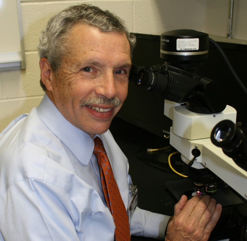
Dr. Perl describes the process by which a brain specimen is studied in the lab.
You obviously don’t keep a count, In my
career I’ve looked at over 20,000 brains. The specimen comes in and it’s examined
with care, it’s photographed, it’s measured, it’s dissected so that various portions
of the brain can be looked at and then actual pieces are prepared so that we can look at
them under the microscope with a whole variety of different types of staining procedures
so that we can look at specific proteins and cell types. It’s a complicated and expensive operation. But at the end of the day one can really detail
the nature of abnormalities present within that brain and can compare it to individuals
who have not been, in this example, exposed to military battlefield conditions to try
to figure out exactly what’s happening.
Posted on BrainLine December 13, 2017.
About the author: Daniel P. Perl, MD
Dr. Perl is a Professor of Pathology at USUHS and Director of the CNRM's Brain Tissue Repository, where he has established a state-of-the-art neuropathology laboratory dedicated to research on the acute and long-term effects of traumatic brain injury among military personnel.