Findings from Structural MR Imaging in Military Traumatic Brain Injury
Riedy G, et al "Findings from structural MR imaging in military traumatic brain injury" Radiology 2015; DOI: 10.1148/radiol.2015150438.
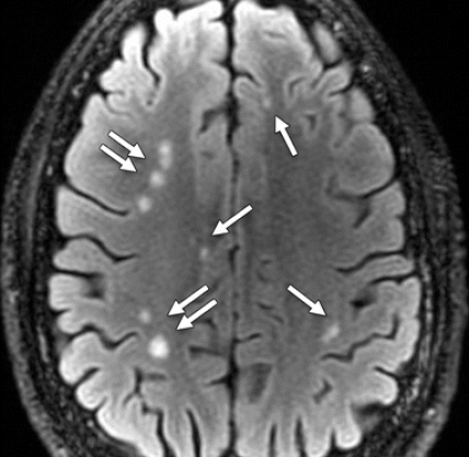
Researchers from the National Capital Neuroimaging Consortium, the National Intrepid Center of Excellence, the Center for Neuroscience and Regenerative Medicine, the Uniformed Services University of the Health Sciences, the Henry M. Jackson Foundation, and Walter Reed National Military Medical Center, in Bethesda, MD, sought to describe initial neuroradiology findings among members of the military service who had experienced a primary chronic mild TBI (MTBI) caused by a blast. Blast-related injury and loss of consciousness is common in military TBI. Structural MR imaging demonstrates a high incidence of white matter T2-weighted hyperintense areas and pituitary abnormalities, with a low incidence of microhemorrhage in the chronic phase.